With lung cancer causing nearly 35,000 deaths in the UK each year[i], the National Optimal Lung Cancer Pathway (NOLCP)[ii,iii] aims to improve outcomes by expediting lung cancer diagnosis. Developed by NHS England, it sets out tight timeframes for each stage of a best practice care pathway.
While this pathway is specifically designed to accelerate detection of lung cancer cases, it sometimes helps to hasten detection of other pathologies that may be equally critical.
The introduction of Annalise Enterprise CXR[iv] has strengthened the system, allowing even the most subtle findings to be detected. In one instance, this occurred within just hours of the solution being deployed in the care pathway.
Understanding the Care Pathway
The standard care pathway at Trusts ensures a streamlined process for patients referred by GPs. Here’s how it works:
- Patient referred by GP presents a request form.
- The imaging request is justified and the x-ray acquired by a radiographer.
- The acquiring radiography team conducts preliminary clinical evaluation (PCE) of the images.
What happens next depends on the preliminary findings.
- If the CXR looks normal on initial review, the patient is sent home to await the formal imaging report.
- If potential abnormalities are identified, the radiographer flags the case for further review by a reporting radiographer or radiologist accelerating the care pathway.
Case Presentation
A male adult in his 20s presented to the North Tees and Hartlepool NHS Foundation Trust hospital with a history of persistent cough and underwent a chest radiograph (CXR) as requested by his GP. The acquiring radiographers did not identify any abnormalities during their PCE. The patient was sent home, consistent with existing protocols.
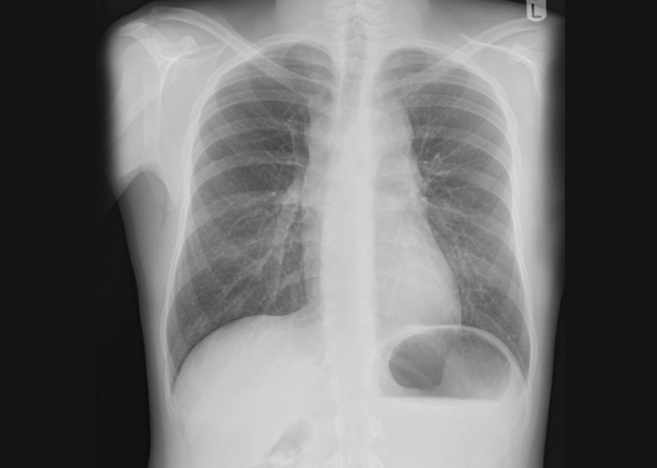
Image 1. Initial CXR of a male adult with persistent cough
Remarkable Timing for a Seemingly Unremarkable Case
Just minutes before this patient’s CXR was taken, the Annalise. ai team were in the process of activating Annalise Enterprise CXR at the site, having completed all testing. The solution had started prioritising cases based on the severity of findings, pushing critical cases to the top of the reporting radiographer’s worklist.
Shortly after the patient’s departure and with Annalise Enterprise now enabled, the reporting radiographer spotted this specific case at the top of the reporting worklist—with a critical priority flagged by the AI.
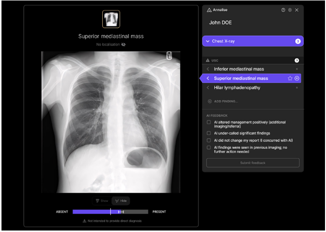
Image 2. Patient CXR with Annalise Enterprise CXR findings
Annalise Enterprise CXR identified 3 critical findings, including hilar lymphadenopathy, superior mediastinal mass and inferior mediastinal mass.
The reporting radiographer immediately reviewed the flagged case and agreed with Annalise Enterprise CXR’s findings, scheduling the patient’s CT scan for the next day. The radiographic appearances indicated a range of potential diagnoses including lymphoma, a significant concern that necessitated urgent follow-up. This change in workflow meant that instead of waiting for reporting in a chronological order (which could take up to a few days depending on staffing levels and CXR volumes), the patient’s images were reported almost immediately. This rapid response helped to ensure timely diagnosis and intervention, potentially saving days on the patient’s pathway.
Follow Up Action and Patient Management
Step 1
The patient underwent a CT scan the following day. A multilocular cystic mass was identified on CT in the mediastinum, accompanied by numerous enlarged lymph nodes located centrally in the chest.
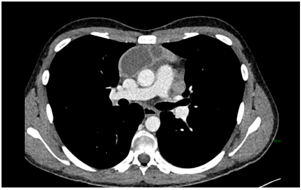
Image 3. CT chest results
The differential diagnosis at this stage included:
- Lymphoma
- Tuberculosis (TB)
- Germ cell tumor (GCT)
- Thymoma
Step 2
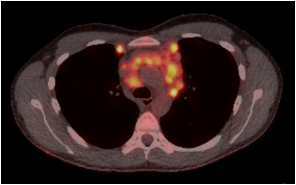
Image 4. PET CT results
Lymphoma was considered the most likely clinical diagnosis and the patient was immediately referred to the appropriate tertiary centre to undergo tissue sampling.
Annalise.ai: Improving Workflows and Patient Outcomes
Implementing an AI-enhanced workflow had an immediate positive impact on departmental efficiency and patient outcomes.

Improved Patient Outcomes and Reduced Delays
Relying solely on chronological report sequences could lead to delayed investigation and treatment, potentially affecting cancer staging, treatment options, and overall prognosis. The introduction of AI reduces this risk by expediting the diagnostic process.

Worklist Prioritisation
Even in a department with an established same-day CT pathway, worklist prioritisation proved immediately valuable, helping ensure that a critical priority case received timely attention. Worklist triage helps draw the attention of acquiring radiographers to prioritise potential cases of clinical significance, ensuring more subtle presentations are not overlooked. This minimises the risk of missed findings, thereby improving diagnostic accuracy.

Enhanced Workflows Help National Standards to be Met
The integration of an AI-enhanced workflow proved instrumental in the timely detection and follow-up for this patient.

[i] Cancer Research UK. Lung cancer statistics. Cancer Research UK. https://www.cancerresearchuk.org/health-professional/cancer-statistics/statistics-bycancer-type/lung-cancer.
[ii,iii] NOLCP Implementation Guide. https://www.roycastle.org/app/uploads/2019/07/Lung_Cancer_Implementation_Guide_August_2017.pdf.
[iv] S oR news article. https://www.sor.org/news/x-ray/teesside-hospital-rolls-out-artificial-intelligenc